The clinical and financial impact of patient motion during MRI
How Siemens MRI technology helps you overcome it

With our award-winning FREEZEit – featuring TWIST-VIBE and StarVIBE – you will be able to take body imaging to the next level by overcoming challenging motion in liver imaging. This will enable you to expand your MRI services in the field of body imaging.
Frost & Sullivan recognizes FREEZEit with the Product Leadership Award for Innovative Techniques in Abdominal Imaging.
StarVIBE

Dynamic liver exams can be challenging for patients, including children, the elderly, and the very sick, who cannot hold their breath for the required time or at all.
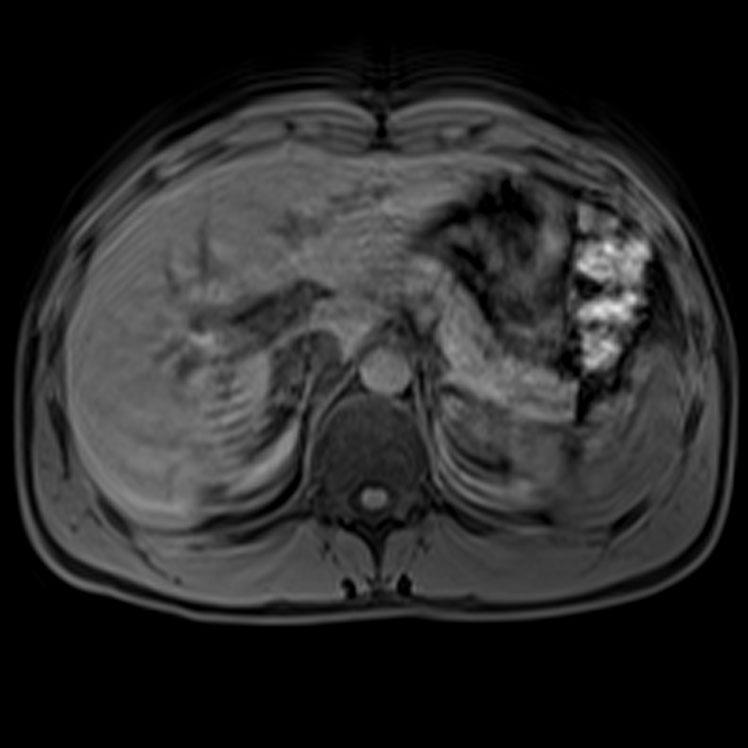
StarVIBE now delivers robust, free-breathing, and contrast-enhanced exams for these patients by intelligently resisting motion artifacts.
Body MRI Case with FREEZEit – Hepatoblastoma Follow-up exam with StarVIBE
Key benefits:
- Reliable imaging for a growing patient population, otherwise excluded from MRI
- Improved treatment due to more accurate results
- More satisfying patient experience
- Less time for preparation
- No need for rescans
“Radial VIBE (StarVIBE) sequence is ideal for MR imaging of pediatric patients. It significantly improves overall image quality and is motion robust, allowing for optimal imaging in free-breathing sedated pediatric patients.“1,2
Sarah S. Milla, MD
Pediatric Radiologist
Atlanta, GA
TWIST-VIBE
Dynamic liver imaging remains a contrast-enhanced technique. Using a contrast agent makes it necessary to catch the right point of the arterial phase. If this point in time is missed, crucial arterial information within the liver remains unseen.
TWIST-VIBE offers high temporal and spatial resolution with full 4D coverage for multi-arterial imaging with 100% precise contrast timing.
Body MRI Case with FREEZEit – FNH/Adenoma Distinction with TWIST-VIBE
Key benefits:
- Robust and fast liver imaging with full 4D coverage
- Excellent images to plan surgical intervention
- Reliable imaging from the very first shot
- Time and cost savings
- No need for rescans
“TWIST-VIBE revolutionizes the diagnostic possibilities for oncologic imaging. It allows for robust, high temporal and spatial resolution scans, improving lesion detection and characterization. Thus, the detection of otherwise almost invisible liver metastases as well as small HCC foci becomes feasible. This immediately impacts further treatment decisions for our patients.”1
Ulrike I. Attenberger, MD
University Medical Center Mannheim
Mannheim, Germany
LiverLab
LiverLab provides non-invasive fat and iron evaluation, and enables for trending and/or monitoring of patients suspicious of liver diseases, especially in early disease stages.
The iron and fat values of the liver are important indicators for a variety of serious illnesses. Early evaluation could be a decisive step to better monitor early stages of diffuse liver diseases such as steatosis and hemochromatosis.
LiverLab is robust and fast enough to be implemented in routine clinical imaging with only a few clicks.
Your benefits:
• Quantitative liver evaluation
• Robust, clinical, inline workflow, built into the Abdomen Dot Engine
• First Look Dixon:
o Single breath-hold (in, out, water, fat)
o Inline, whole liver segmentation
o Inline liver volume calculation
o Evaluation suggestion: if fat / iron is detected


from “First Look” Dixon
Patient with focal iron deposition
- Multi-Echo Dixon
o Inline parametric maps of the whole liver
- T2*, R2*
- Fat and water fraction
o Inline evaluation and reports
- Color bar charts
- Parameter values over the specified ROI
- Parameter values and histograms of the whole liver


Patient with relatively severe fatty liver
- HISTO
o Multi-echo single voxel spectroscopy
o User control of voxel placement
o Inline evaluation and reports
- Color bar chart
- Spectra and fitting curves for quality control
- Fat fraction
- R2 water

CAIPIRINHA (Controlled Aliasing in Parallel Imaging Results in Higher Acceleration)
Cutting your 3D breath-holds in half
With the new CAIPIRINHA (Controlled Aliasing in Parallel Imaging Results in Higher Acceleration) technique, you can accelerate acquisition and significantly reduce 3D breath-holds—without affecting image resolution, coverage, or contrast.
CAIPIRINHA acquisition can be used with 3D TI VIBE or 3D Dixon technique—which provides four contrasts (in phase, opposed phase, fat, and water) in a single short breath-hold.
25 second breath hold without CAIPIRINHA
3D VIBE FS
3mm, 256 matrix, 72 slices

7 second breath hold with CAIPIRINHA
3D VIBE FS CAIPIRINHA x 4
3mm, 288 matrix, 72 slices

3D Dixon Water CAIPIRINHA x 4
3mm, 320 matrix, 64 slices

3D Dixon FAT CAIPIRINHA x 4
3 mm, 320 matrix, 64 slices

3D Dixon In Phase CAIPIRINHA x 4
3mm, 320 matrix, 64 slices

3D Dixon Opposed Phase CAIPIRINHA x 4
3mm, 320 matrix, 64 slices
CAIPIRINHA software is included as standard with Siemens’ D13A software for MAGNETOM Aera, Skyra, Avanto and Verio. It is also available with D14 software for the MAGNETOM ESSENZA 1.5T system.
Decrease breath holds – increase workflow
Abdominal MRIs can be challenging—motion (voluntary and involuntary), breath-hold capacity of the patient, acquisition speed, and contrast timing can all impact body imaging. If you could improve your abdominal MRIs, you can yield significant benefits.
Improve your abdominal MRI service and you’ll be able to:
- Increase patient compliance
- Enhance patient satisfaction
- Have consistency among technologists
- Reduce repeat exams, increase throughput
- Improve image quality and diagnostic confidence
- Attract more referrals
- Meet utilization goals
“With CAIPIRINHA, acquisition times shorter than 10 seconds could have been achieved, which is a tremendous advantage, especially for elderly patients, who are not able to suspend their breath sufficiently. […] CAIPIRINHA-accelerated T1w-VIBE imaging seems to be a very robust and reliable technique. At our institution, this technique is now the clinical standard for abdominal MR studies.”
Highly Accelerated T1-Weighted Abdominal Imaging Using 2-Dimensional Controlled Aliasing in Parallel
Imaging Results in Higher Acceleration.
Riffel P, Attenberger UI, et. al.
Investigative Radiology. Volume 48, Number 7, July 2013.
Abstract
Controlled Aliasing in Parallel Imaging Results in Higher Acceleration (CAIPIRINHA)
Download CAIPIRINHA Flash article
Elastography
Elastography
Liver fibrosis with its associated cirrhosis and hypertension is a highly prevalent disease in the U.S. and traditionally requires biopsy for diagnosis. MR Elastography (MRE) offers the possibility to evaluate relative liver stiffness noninvasively.
Wave Images
2D GRE MRE sequence Is run during application of mechanical waves by active / passive drivers. Wave images allow visualization of resulting shear waves as they travel through the tissue

Inline Elastogram
Calculated from wave image provides data about relative tissue stiffness. Here red indicates higher stiffness in fibrotic liver.

Elastogram with Confidence Mask1
Siemens unique. Provides criteria to determine if calculated sheer wave stiffness values for a specific voxel is reliable
Our MRE package includes the hardware and software features to incorporate this state-of-the-art diagnostic tool into your diagnostic MR abdominal service. With it, you receive:
- Active driver
- Passive driver
- Body 18 coil (standard)
- 2D gradient echo sequence with
- Motion-Encoding Gradients
- iPAT enabled for shortened breath-holds
- Inline Elastogram
- Confidence Mask1
- Magnitude, Color Stiffness, Color Stiffness with Confidence Mask and Color Wave images ready to read
- Ability to reading stiffness directly on the color elastogram1
- Free window leveling of color elastograms1


